 What Are FFPE Tissue Sections?
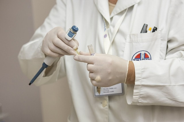
Unless you are in the medical line or profession, the term FFPE will be strange indeed. But for those in the medical field, FFPE Tissue Sections (Formalin-Fixed Paraffin-Embedded Tissues) is one of the most critical and important tools for research and treatment of some of the most problematic diseases afflicting mankind. It is being used in research of complex diseases ranging from cancer to Alzheimer's disease. By description, FFPE tissues are biopsy specimen that have been preserved in formaldehyde, otherwise known as formalin. These samples are what pathologists work with to properly diagnose certain ailments like the ones stated above. What, And How Is FFPE tissues Used? Immunohistochemistry (IHC) is the science of placing a biopsy (thin slice of tissue) specimen, which are bathed in certain solutions to preserve the molecular structures, under a microscope in order to study or accentuate molecular structural deformities. The type of protein structure, and/or deformity shown will determine the disease, be it cancer, Alzheimer's disease, including some other forms of maladies. This type of procedure is very common with cancer, and other diseases' research. The critical aim is to study the structure of molecules and proteins in these tissues. FFPE Tissue Sections, Preparation And Storage For research purposes, human tissue may be taken from a donor or patient, primates, snakes and of course, lab mice also offer good blocks of tissues. The size and thickness of the tissue cut or excised is usually a function of the research purpose, and the nature of the disease to be analized. The thickness of tissues used usually measure about 1*1*0.5 cubic centimeters, although this may vary, but generally the size is within this range. The tissues are then immersed in a 10 % solution of formalin that is of neutral buffer for a period of between 18 hours and, but not more than 24 hours. This is then finally embedded into IHC-grade paraffin. Properly prepared FFPE tissue sections are quite durable and can be stored even at ordinary room temperature. Therapeutic Uses Of FFPE Tissue Sections In The Study Of Oncology > Generally, tumor tissues exhibit some unique morphological characteristics, the use of FFPE Tissue in the study of Oncology has therefore become increasingly invaluable. Therefore, identifying the presence of certain proteins and their structures are used by researchers to determine if and when a therapeutic intervention might, or might not be successful. Immunology > Studying the reaction of the immune system, when in a healthy or diseased state, particularly after taking samples of an autoimmune disease patient, helps in determining the likely outcome of certain interventionist approach to treating autoimmune diseases. Samples From Healthy Donors And Future Research > There's is a great deal of ongoing research, and proposed future work which require well prepared and archived FFPE Tissue Sections as resources for DNA, RNA etc. There are even healthy donors who have willed their bodies to science research in this line, these will eventually become very invaluable for future comparative analysis. Formats (Methods) Of Collection There are a few formats for collection of specimen, this usually includes, 1). matching samples from the same individuals. 2) Healthy or diseased donors are also a valuable source for specimen collection. 3). Non-human source for specimen collection also include: lab mice, primates and snakes.
0 Comments
The validation of an innovative concept on actual human tissue remains a classic issue in biology. Since the first sampling is being conducted by a pathologist there will be a shortage of tissue. This is because the goal of a pathologist is to provide a diagnostic consultation. Pathologists do not provide material for future experimentation. On this note, it is important to deliberate on tissue microarray uses. Reading through the remaining part of the article will help to divulge more tissue microarray uses.
Tissue Retention: The block may be cored in these cases for a few times without damaging them. There is every possibility to perform a diagnosis upon subsequent sectioning. This can even occur if the tissue has been separated for an array-oriented research. Staining: Tissue microarrays are amenable to a plethora of strategies including immunologic stains and histochemical stains, fluorescent or chromogenic visualization. Tissue microarray uses can also be found in situ hybridization, microdissection, and other techniques. This creates a huge difference between microarray tissues and the conventional formalin-fixed paraffin cascaded material. The Durability Of Antigen: Comparing the stains of two to ten microarray disks will help you discover a great misery. The whole tissue sections from which the disks are derived should be tested and analyzed accordingly. It is clear that the analysis or observation of 2 disks is highly comparable than a full tissue section. There is over ninety-five percent accuracy in several cases for this analysis. With two-fold redundancy, the tissue microarray strategy remains an accurate and valuable way for analyzing protein expression in huge archival cohorts. Tissue Conservation: Do the tiny histo-spots represent a whole section? Tissue volume remains the primary restriction of this strategy. Some critics have mentioned that the amount of item used in the analysis may be too small. Skeptics also claim that it may not be a representation of the whole tumor. There is every possibility for any histo-spot to remain negative on a particular array. In the ultimate conclusions, the statistical power of studies removes the chance of variability. This shows that microarray tissues are highly conservative. Tissue Microarray Uses For Immunohistochemical Research Of Ameloblastoma: Ameloblastoma remains a traditional and tough odontogenic tumor. This tumor has a high rate of reoccurrence for real. Tissue microarray can be used as an important tool to understand the molecular function of ameloblastoma. For different human neoplasms, tissue microarrays have been considered a high-throughput strategy. The truth is that it needs to be confirmed in the ameloblastoma study. After several studies and experiments, it is evident that tissue microarray uses can be confirmed by an immunohistochemical study of ameloblastoma. It helps to determine and analyze the most appropriate design. Conclusion: The uses of tissue microarray and a frozen tissue array are endless since more studies on this technique continue to grow. Some experts can also use the TMA technique in testing and quantify cfDNA and other important experiments. Without any scintilla of doubt, it is unequivocal that this technique remains unique and special. It helps to study the longevity of antigen and other important items within tissue sectioning. |
 RSS Feed
